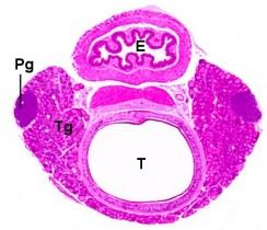
Transverse section.
| Species: | Rats and Mice |
| Organs: | Thyroid gland Parathyroid gland Trachea Esophagus |
| Localization: | In the area of the parathyroid gland |
| Number of sections: | 1 |
| Direction: | If thyroid glands are not weighed: Transverse section of trachea, esophagus, thyroid and parathyroid glands. Optional: longitudinal horizontal section of thyroid glands in conjunction with trachea. A separate transverse section of the esophagus is made. If thyroid glands are weighed: Longitudinal horizontal (largest cut surface), section of thyroid and parathyroid glands. Transverse section of trachea and esophagus. |
| Remarks: | Recuts are sometimes required to consistently include the parathyroid in the section. The number of focal lesions observed depends on the area of thyroid examined. |
 Transverse section. |
 Transverse section (T: trachea, Tg: thyroid gland, Pg: parathyroid gland, E: esophagus). |
 Thyroid gland, longitudinal section, with parathyroid gland (Pg). |
Relevant differences between rats and mice:
The rat possesses only one pair of parathyroids. They are located on the anterior and lateral aspect of the thyroid lobes but may vary in position.
In the mouse, the position and the number of parathyroids is variable. Usually, there are two parathyroid glands located bilaterally just under the capsule near the dorsolateral border of each thyroid lobe. They are rarely found at the same level, sometimes one or both may be posterior to the thyroid; they may be deeply embedded in the thyroid tissue and there may be more than two.
See also:
Trachea (inhalation study)
Esophagus with Trachea
Introduction
Boorman GA, DeLellis RA (1983) C-cell hyperplasia, thyroid, rat. In: Jones TC, Mohr U, Hunt RD (eds): Monographs on pathology of laboratory animals. Endocrine System. Springer, Berlin Heidelberg New York, pp 192–204 | |
Botts S, Capen CC, DeLellis RA, Deschl U, Hartig F, Karbe E, Konishi Y, Krinke GJ, Landes C, Mettler F, Rebel W, Riley MGI, Tuch K, Urwyler H (1994) 6. Endocrine System. In: Mohr U, Capen CC, Dungworth DL, Griesemer RA, Ito N, Turusov VS (eds) International classification of rodent tumours. Part I, The rat. IARC Scientific Publications No. 122, Lyon | |
Botts S, Jokinen NP, Isaacs DJ, Meuten DJ, Tanaka N (1991) Proliferative lesions of the thyroid and parathyroid glands. E-3. Guides for Toxicologic Pathology, STP/ARP/AFIP, Washington | |
Capen CC (1996) Hormonal imbalances and mechanisms of chemical injury of thyroid gland. In: Jones TC, Capen CC, Mohr U (eds) Monographs on pathology of laboratory animals. Endocrine system, 2nd edition. Springer, Berlin Heidelberg New York Tokyo, pp 245–238 | |
Capen CC (1996) Pathobiology of parathyroid structure and function in animals. In: Jones TC, Capen CC, Mohr U (eds) Monographs on pathology of laboratory animals. Endocrine system, 2nd edition. Springer, Berlin Heidelberg New York Tokyo, pp 293–327 | |
Kittel B, Ernst H, Kamino K (1996) Anatomy, histology and ultrastructure, parathyroid, mouse. In: Jones TC, Capen CC, Mohr U (eds) Monographs on pathology of laboratory animals. Endocrine system, 2nd edition. Springer, Berlin Heidelberg New York Tokyo, pp 328–329 | |
Kittel B, Ernst H, Kamino K (1996) Anatomy, histology and ultrastructure, parathyroid, rat. In: Jones TC, Capen CC, Mohr U (eds) Monographs on pathology of laboratory animals. Endocrine system, 2nd edition. Springer, Berlin Heidelberg New York Tokyo, pp 330–332 | |
Pozharisski KM (1990) Tumours of the oesophagus. In: Turusov V, Mohr U (eds) Pathology of tumours in laboratory animals. Vol I. Tumours of the rat, 2nd edition. IARC Scientific Publications No. 99, Lyon, pp 109–128 |
|
Trm V 5.00 |
Reference: Kittel B, Ruehl-Fehlert C, Morawietz G, et al. (2004) Revised guides for organ sampling and trimming in rats and mice – Part 2. A joint publication of the RITA and NACAD groups. Exp Toxic Pathol 55: 413–431 |