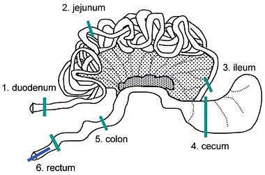
Intestine in situ with mesentery and mesenteric lymph nodes.
| Species: | Rats and Mice |
| Organs: | Small intestine Duodenum Jejunum Ileum Large intestine Cecum Colon Rectum Peyer's patches |
| Localizations: | 1) Duodenum: 1 cm distal to the pyloric sphincter 2) Jejunum: central section 3) Ileum: 1 cm proximal to cecum 4) Cecum 5) Colon: central section 6) Rectum: 2 cm proximal to the anus |
| Number of sections: | 6 |
| Direction: | Transverse |
| Remarks: | Duodenum in conjunction with an adjacent piece of pancreas Jejunum containing Peyer's patch or lymph follicle Optional: additional longitudinal and/or transverse section through Peyer's patches Cecum: due to the large diameter it is advisable to open the specimen Rectum optional: longitudinal vertical section to include the anus |
 Intestine in situ with mesentery and mesenteric lymph nodes. |
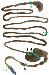 Intestine: for the collection of specimens, the mesentery is removed (1: duodenum, 2: jejunum, 3: ileum, 4: cecum, 5: colon, 6: rectum). |
 Intestine in situ. |
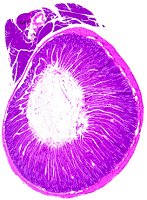 Duodenum. |
 Jejunum (left) and ileum (right). |
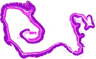 Cecum. |
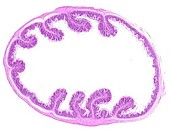 Colon. |
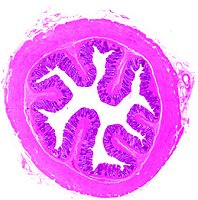 Rectum. |
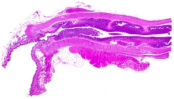 Rectum, longitudinal section (optional). |
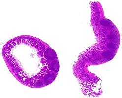 Jejunum with Peyer's patches (optional). |
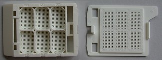 Tissue Tek Cassette, Fa. Vogel, Giessen, Germany. |
During necropsy or after fixation, the intestine is carefully separated from the mesentery. Peyer's patches of the jejunum are mostly visible as slightly elevated lighter fields in the intestine's wall or are even discernible as prominent areas when activated. One transverse section from each part of the unopened bowel is taken. The remaining intestine should be opened and examined for abnormalities. At necropsy, the ingesta should not be removed vigorously but only gently rinsed with physiological saline if necessary.
Swiss roll technique: This technique is sometimes required for examination of the whole intestine and the gut associated lymphatic tissue (GALT). The intestine is stripped off the mesentery, opened with a pair of scissors and gently rinsed. The intestine except cecum is recoiled on cotton swabs and fixed. After fixation, the spooled intestine is detached and embedded. This procedure is technically challenging and not recommended for routine purposes, as the intestinal mucosa and the lymph follicles will often be found cut tangential. However, transverse sections as described above will often provide a better histoanatomy.
The jejunum and ileum or the distal colon and rectum cannot readily be differentiated microscopically. For consistency in routine examination of each required site, accurate sampling is necessary. In this case, the colon differs from the rectum by a thinner muscle layer and a larger lumen. For dehydration and embedding, cassettes with a subdivision are helpful.
Please note that the magnification of the histological images is not the same for all parts of the intestine.
See also:
Pancreas
Introduction
|
Trm V 5.00 |
Reference: Ruehl-Fehlert C, Kittel B, Morawietz G, et al. (2003) Revised guides for organ sampling and trimming in rats and mice – Part 1. A joint publication of the RITA and NACAD groups. Exp Toxic Pathol 55: 91–106 |