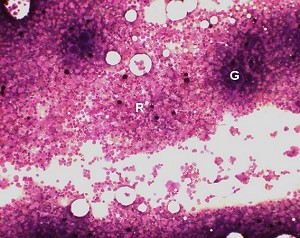
Low magnification of a rat bone marrow smear, stained with May-Gruenwald-Giemsa, showing grossly visible particles (G) and regions suitable for evaluation (R).
| Species: | Rats and Mice |
| Organ: | Bone marrow |
| Localization: | Core sample from the femoral diaphysis |
| Number of sections: | 1 |
| Remarks: | Slides should only be stored after fixation in methanol. Staining with the Pappenheim, May-Gruenwald-Giemsa or Wright method. |
 Low magnification of a rat bone marrow smear, stained with May-Gruenwald-Giemsa, showing grossly visible particles (G) and regions suitable for evaluation (R). |
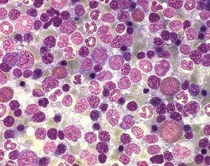
 High power view of a rat bone marrow smear stained with May-Gruenwald-Giemsa. |
Various techniques have been described. For each method training is necessary to obtain satisfactory smears. Femur diaphysis is recommended as localization because no bone trabecula are intermingled with the hematopoietic tissue.
The proximal and distal epiphyses are cut off with scissors. For removal of the bone marrow, air is blown from one end into the marrow cavity and the marrow cast is collected onto a glass slide. The smear is prepared conventionally with a cover glass. A smear of adequate quality contains grossly visible particles. Marrow smears should be prepared as fresh as possible to avoid blood clotting.
For collection of the bone marrow, aspiration with a pipette containing anticoagulated serum or a small paint brush or a cotton bud from the longitudinally opened femur can also be used.
See also:
Introduction
Valli VE, McGrath JP, Chu I (2002) Hematopoietic system. In: Haschek WM, Rousseaux CG, Wallig MA (eds) Handbook of toxicologic pathology, 2nd edition, Vol 2. Academic Press, San Diego New York Boston, pp 647–679 | |
Valli VE, Vileneuve DC, Reed B, Barsoum N, Smith G (1990) Evaluation of blood and bone marrow, rat. In: Jones TC, Ward JM, Mohr U, Hunt RD (eds) Monographs on pathology of laboratory animals. Hemopoietic system. Springer, Berlin Heidelberg New York Tokyo, pp 9–26 |
Trm V 5.00 |
Reference: Morawietz G, Ruehl-Fehlert C, Kittel B, et al. (2004) Revised guides for organ sampling and trimming in rats and mice – Part 3. A joint publication of the RITA and NACAD groups. Exp Toxic Pathol 55: 433–449 |