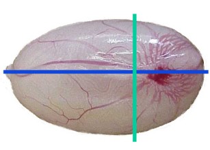
Testis, recommended transverse section (green), optional section (blue).
| Species: | Rats and Mice |
| Organs: | Testis Rete testis |
| Localization: | Transverse, close to the rete testis |
| Number of sections: | 2 (1 per side) |
| Direction: | Transverse |
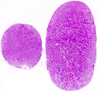
| Remarks: | A transverse section containing the area of the rete testis provides good histology of seminiferous tubules as well as of the rete testis. Optional: longitudinal vertical section containing the rete testis. |
 Testis, recommended transverse section (green), optional section (blue). |
 Testis, transverse section left, optional longitudinal section right. |
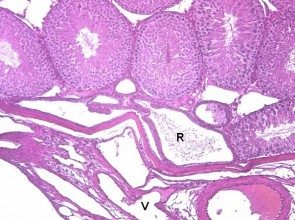
 Rete testis (R), rat (V: vasculature). |
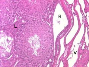
 Rete testis (R), mouse (V: vasculature, L: Leydig cells). |
Near the capsule, focal tubules with flat or incomplete epithelium can be found. These should not be confused with atrophic or degenerating tubules, as they represent tubules of the rete testis. Generally, the rete testis is very small in rodents and not well visible in the histological section. In mice it is slightly more prominent than in rats and hyperplasia of the rete testis can be observed in older animals. For inclusion of the rete testis in the histological section orientation is given by the vasculature.
In short-term studies, fixation with Davidson's or Bouin's solutions is highly recommended to detect less extensive toxicity.
Leydig cells (interstitial cells) are present in small groups in the interstitium between the seminiferous tubules. They are found in a similar distribution in all sections proposed.
See also:
Introduction
|
Trm V 5.00 |
Reference: Kittel B, Ruehl-Fehlert C, Morawietz G, et al. (2004) Revised guides for organ sampling and trimming in rats and mice – Part 2. A joint publication of the RITA and NACAD groups. Exp Toxic Pathol 55: 413–431 |