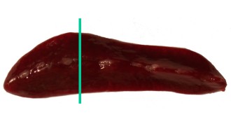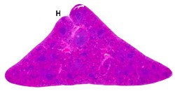
Spleen.
| Species: | Rats and Mice |
| Organ: | Spleen |
| Localizations: | At largest extension Option: whole organ |
| Number of sections: | 1 |
| Direction: | Transverse Option: longitudinal horizontal (not shown in the image) |
 Spleen. |
 Spleen (H: hilus). |
A transverse section is made at the largest extension of the organ, showing red and white pulp. This plane of section guarantees the presence of all relevant anatomical structures and hallmarks of the white pulp, e.g. PALS (periarteriolar lymphatic sheath), marginal zone and follicles.
See also:
Introduction
|
Trm V 5.00 |
Reference: Morawietz G, Ruehl-Fehlert C, Kittel B, et al. (2004) Revised guides for organ sampling and trimming in rats and mice – Part 3. A joint publication of the RITA and NACAD groups. Exp Toxic Pathol 55: 433–449 |