Spinal cord.
| Species: | Rats and Mice |
| Organs: | Spinal cord Spinal nerve root |
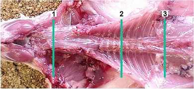
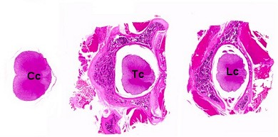
| Localizations: | 1) Cervical cord at upper cervical segment. Optional: cervical cord cut at the level of the first cervical nerve (see brain) 2) Thoracic cord at mid-thoracic segment 3) Lumbar cord at 4th lumbar segment close to last rib |
| Number of sections: | 3 |
| Direction: | Transverse |
| Remarks: | In conjunction with vertebral body (decalcified). Optional: without bone to avoid decalcification. Embedded with anterior faces down. |
 Spinal cord. |
 Spinal cord (Cc: cervical cord, Tc: thoracic cord, Lc: lumbar cord). Option without bone shown for the cervical cord. |
Transverse sections of the spinal cord are required to assess whether the findings are distributed uni- or bilaterally, and symmetrically or asymmetrically. Sections should be obtained at the upper cervical, mid-thoracic and lumbar levels. The upper cervical area positioned at the transition of medulla oblongata to cervical spinal cord (level of the first cervical nerve) is important for the detection of distally accentuated neuropathy affecting the long spinal tracts. This region is known to produce the earliest and most prominent lesions in the dorsal cervical column. At necropsy, the spinal cord should be cut off directly posterior to the cerebellum and can be removed with the vertebral bone as with the thoracic and lumbar spinal cord.
In the lumbar spinal cord, the area of L4 segment should preferably be examined, as this segment provides the main contribution to the peripheral sciatic nerve. The L4 segment of the spinal cord is located at the junction of the thoracic and lumbar spine which is indicated by the last rib. If the lumbar specimen is taken more caudally, it will contain only the cauda equina.
It is advantageous to remove the spinal cord from the vertebrae before processing to avoid decalcification. However, processing in situ has the advantage to include dorsal root ganglia and nerve roots to assess radiculoneuropathy.
In neurotoxicity studies, sections from the cervical and lumbar swellings are recommended in current guidelines (OECD 424). The cervical swelling is located at the C3 - C6 segments of the spinal cord. This section is not prioritized but can be taken as additional option in routine rodent studies.
To avoid artifactual vacuolation in the white matter, the specimens should not be stored in alcohol.
See also:
Brain
Introduction
Gelderd JB, Chopin SF (1977) The vertebral level of origin of spinal nerves in the rat. Anat Rec 188: 45–48 | |
Krinke GJ (1989) Neuropathologic screening in rodent and other species. J Am Coll Toxicol 8: 141–146 | |
Krinke GJ, Fitzgerald R (1988) The pattern of pyridoxine-induced lesion: difference between the high and the low toxic level. Toxicology 49: 171–178 | |
OECD (1997) Test guideline 424. Neurotoxicity study in rodents | |
Schäppi U, Krinke G (1991) Die Erfassung toxischer Wirkungen am Nervensystem. In: Hess R (ed) Arzneimittel-Toxikologie. Georg Thieme Verlag, Stuttgart, New York, pp 238–258 |
|
Trm V 5.00 |
Reference: Morawietz G, Ruehl-Fehlert C, Kittel B, et al. (2004) Revised guides for organ sampling and trimming in rats and mice – Part 3. A joint publication of the RITA and NACAD groups. Exp Toxic Pathol 55: 433–449 |