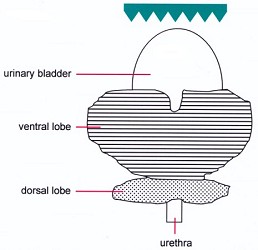
Prostate, rat, ventral aspect. Lateral lobes not visible, dorsal lobe: only caudal part visible.
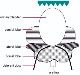
Prostate, rat, dorsal aspect.
| Species: | Rats and Mice |
| Organ: | Prostate |
| Localization: | Dorsolateral and ventral lobe |
| Number of sections: | 1 |
| Direction: | Longitudinal horizontal after special preparation (see below). |
 Prostate, rat, ventral aspect. Lateral lobes not visible, dorsal lobe: only caudal part visible. |
 Prostate, rat, dorsal aspect. |
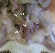 Rat, abdominal cavity, ventral aspect. In situ localization of prostate and attached organs. |
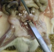 The urethra is dissected. |
 Removal of prostate, urinary bladder, and seminal vesicles as a unit. |
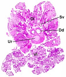 Prostate. |
Abbreviations used in the above images: Sv: seminal vesicle, Cg: coagulation gland, Ub: urinary bladder, Ur: urethra, Vl: ventral lobe of prostate, Dl: dorsolateral lobe of prostate, Dsv: duct of seminal vesicle, Dd: deferent duct
The dorsolateral and ventral lobes that normally lie in a vertical axis above each other (with urinary bladder and seminal vesicle in between) are spread in a horizontal axis and embedded with the "outer" aspect down into the cassette.
Preparation: The group of adjacent organs consisting of prostate, urinary bladder, seminal vesicles and coagulation glands is removed (see figures 4.4.d through 4.4.f) and (if weights are not required) fixed in toto to prevent leakage of the glandular secretions.
After fixation, the ventral lobe is detached from the urinary bladder and is flipped back. The urinary bladder and seminal vesicles with coagulation glands are removed. The two ventral lobes are separated from each other, but are left attached to the dorsolateral parts. The dissected prostate is put into a cassette with the "outer" surfaces down; i.e. ventral face of the ventral lobes down and dorsal face of the dorsolateral lobes down (see figures 4.4.g through 4.4.i). After histotechnical processing, a section at the mid level of the ventral lobes is made.
The dorsocranial lobe of the prostate (i.e. coagulating gland) is processed with the seminal vesicle.
Chemically induced or spontaneous proliferative lesions of the rat prostate can be found in all three lobes. The dorsal and lateral lobes exhibit the same spectrum of proliferative lesions. These differ from spontaneous and induced lesions in the ventral lobe. Additionally, some strain specific deviations in the interlobular distribution of benign and malignant neoplasms consequently require the assessment of all compartments. Accordingly, a longitudinal-horizontal section through the prostate complex, including dorsolateral and ventral lobes, urethra and, optionally, ureter and ductus deferens represents a less time consuming method, applicable to routine histological processing and examination.
See also:
Seminal vesicle and coagulating gland
Introduction
|
Trm V 5.00 |
Reference: Kittel B, Ruehl-Fehlert C, Morawietz G, et al. (2004) Revised guides for organ sampling and trimming in rats and mice – Part 2. A joint publication of the RITA and NACAD groups. Exp Toxic Pathol 55: 413–431 |