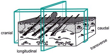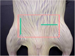
Skin, transverse and longitudinal cutting direction.

Skin, inguinal region: transverse and longitudinal cutting direction.
| Species: | Rats and Mice |
| Organs: | Mammary gland Skin/subcutaneous tissue |
| Localization: | Inguinal region |
| Number of sections: | 1 |
| Direction: | a) Transverse b) Longitudinal vertical to the direction of the hair flow |
| Sample size: | 1 x 3 cm |
| Remarks: | Transverse section: includes the nipple and the lateral iliac lymph node. Longitudinal section: the nipple is not included if the lymph node is enclosed. Both sections: ensure a high amount of mammary gland tissue. |
 Skin, transverse and longitudinal cutting direction. |
 Skin, inguinal region: transverse and longitudinal cutting direction. |
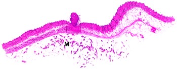
 Skin and mammary gland (M), section transverse to the direction of the hair flow. |
 Skin, longitudinal section in the direction of the hair flow. |
The mammary gland is a paired organ. Due to the diffuse distribution of mammary gland tissue it is of no concern whether one or both sides are in the section. The inguinal region is the recommended area for harvesting mammary gland. Sections of mammary gland should be taken with associated nipple and skin. The result of histotechnique may be improved by shaving the skin at necropsy or removing the hair with scissors at trimming. Orientation of a shaved skin specimen is possible by the nipples in female animals. In male animals, the inguinal region is also preferred to examine skin and mammary gland tissue. The section will be embedded on the cut edge so that it reveals skin, subcutis and mammary gland close to the nipple. In the longitudinal section, the hair follicles will be visible in full length.
Relevant differences between rats and mice
Rats have 6 pairs of mammary glands while mice have only 5 pairs. This difference is not of practical importance since mammary tissue is abundant in the inguinal region of both species. In females, the mammary tissue extends from the salivary gland region to the base of the tail.
See also:
Introduction
|
Trm V 5.00 |
Reference: Ruehl-Fehlert C, Kittel B, Morawietz G, et al. (2003) Revised guides for organ sampling and trimming in rats and mice – Part 1. A joint publication of the RITA and NACAD groups. Exp Toxic Pathol 55: 91–106 |