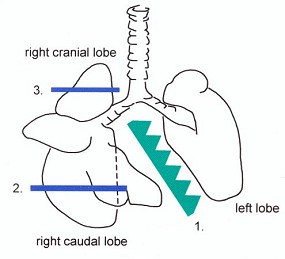
Lung, ventral aspect, oral study.
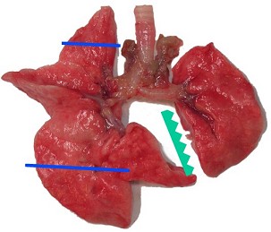
Lung, ventral aspect, oral study.
| Species: | Rats and Mice |
| Organs: | Lung Bronchus Bronchiole |
| Localizations: | Recommended procedure: 1) Left lobe 2) Optional: right caudal lobe 3) Optional: right cranial lobe |
| Number of sections: | 1 (3) |
| Direction: | Longitudinal horizontal Optional: transverse |
| Remarks: | Instillation strongly recommended. Sectioning to the axis of the lobar bronchus. Longitudinal section comprising the lobar bronchus and its main branches. Sample size(s) adapted to the size of the cassette(s). Alternative procedure: Rat: right lobes embedded ventral surface down. Mouse: whole lung embedded, ventral surface down. |
 Lung, ventral aspect, oral study. |
 Lung, ventral aspect, oral study. |
| Species: | Rats |
| Localizations: | 1) Left lobe 2) Right caudal lobe 3) Right cranial lobe 4) Right middle lobe 5) Accessory lobe |
| Number of sections: | 5 |
| Direction: | Sections 1, 2: longitudinal horizontal Sections 3, 5: transverse Section 4: longitudinal vertical |
| Remarks: | Instillation obligatory. Longitudinal horizontal section comprising the lobar bronchus and its main branches. Sample size(s) adapted to the size of the cassette(s); preferentially, the diaphragmatic margin is trimmed off. Alternative procedure: right and left lobes (separate blocks) embedded ventral surface down. |
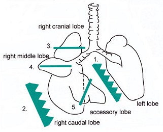 Lung, rat, ventral aspect, inhalation study. |
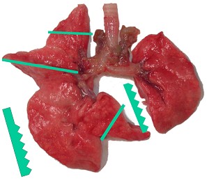 Lung, rat, ventral aspect, inhalation study. |
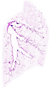 Lung, rat, location 1, left lobe. |
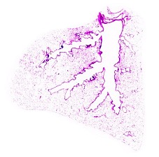 Lung, rat, location 2, right caudal lobe. |
 Lung, rat, location 3, right cranial lobe. |
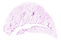 Lung, rat, location 4, right middle lobe. |
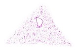 Lung, rat, location 5, accessory lobe. |
| Species: | Mice |
| Localizations: | 1) Left lobe 2) Right caudal lobe 3) Right cranial lobe 4) Right middle lobe 5) Accessory lobe |
| Number of sections: | 5 |
| Direction: | Sections 1, 2, 4, 5: longitudinal horizontal Section 3: transverse |
| Remarks: | Instillation obligatory. Similar procedure as in rats, but lobes are embedded in toto, ventral surface down and detached from the trachea. The five lobes normally fit into one cassette. Option: whole lung in toto (ventral surface down) without removal of the trachea. Microtome sectioning of left lobe and right caudal lobe until lobar bronchus and its main branches are visible (longitudinal-horizontal axis). |
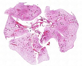 Lung, mouse, in toto (option). |
Rats and mice:
Spontaneous neoplastic pulmonary lesions are rare in rats and arise mostly in the lung periphery whereas regenerative hyperplasia and squamous metaplasia occur mainly in the centroacinar region. Therefore tissue of the lung including parenchyma, bronchiolo-alveolar junctions and main bronchi should be investigated. In oral toxicity studies, at minimum, one longitudinal section of the left lobe should be examined. Additionally, transverse sections of the right cranial and caudal lobes may be examined. In these sections, the epithelium of the major bronchioles, which is one important site of lesions, can be examined at its widest diameter. In inhalation studies, sections of all five lobes should be examined according to the proposed scheme, which facilitates unambiguous identification of individual lung lobes. For histological identification of proliferative lesions in the lung, careful fixation by intratracheal instillation is recommended, even for oral studies.
See also:
Introduction
Dungworth DL, Ernst H, Nolte T, Mohr U (1992) Nonneoplastic lesions in the lungs. In: Mohr U, Dungworth DL, Capen CC (eds) Pathobiology of the aging rat. Vol I. ILSI Press, Washington, pp 143–160 | |
Gopinath C, Prentice DE, Lewis DJ (1987) The respiratory system. In: Atlas of experimental toxicological pathology. MTP Press, Lancaster, Boston, The Hague, pp 22–42 | |
Plopper CG (1996) Structure and function of the lung. In: Jones TC, Dungworth DL, Mohr U (eds) Monographs on pathology of laboratory animals. Respiratory system, 2nd edition. Springer, Berlin Heidelberg New York Tokyo, pp 135–150 | |
Renne R, Fouillet X, Maurer J, Assaad A, Morgan K, Hahn F, Nikula K, Gillet N, Copley M (2001) Recommendation of optimal method for formalin fixation of rodent lungs in routine toxicology studies. Toxicol Pathol 29: 587–589 | |
Rittinghausen S, Dungworth DL, Ernst H, et al. (1992) Primary pulmonary tumours. In: Mohr U, Dungworth DL, Capen CC (eds) Pathobiology of the aging rat. Vol I. ILSI Press, Washington, pp 161–172 | |
Schwartz LW, Hahn FF, Keenan KP, et al. (1991) Proliferative lesions of the rat respiratory tract. R-1. In: Guides for toxicologic pathology. STP/ARP/AFIP, Washington | |
Sminia T, van der Brugge-Gamelkoorn G, van der Ende MB (1990) Bronchus-associated lymphoid tissue, rat, normal structure. In: Jones TC, Ward JM, Mohr U, Hunt RD (eds) Monographs on pathology of laboratory animals. Hemopoietic system. Springer, Berlin Heidelberg New York Tokyo, pp 300–307 |
|
Trm V 5.00 |
Reference: Kittel B, Ruehl-Fehlert C, Morawietz G, et al. (2004) Revised guides for organ sampling and trimming in rats and mice – Part 2. A joint publication of the RITA and NACAD groups. Exp Toxic Pathol 55: 413–431 |