Kidney and ureter.

Kidney and ureter (U) in situ.
| Species: | Rats and Mice |
| Organs: | Kidney Renal pelvis Ureter |
| Localizations: | Kidney: both in the median, through the tip of papilla and renal pelvis. Ureter: transverse section midway between kidneys and bladder. Optional: adjacent to the renal pelvis (not shown in the image). |
| Number of sections: | 2 (1 per side) |
| Direction: | Kidney: one side longitudinal, other side transverse Ureter: longitudinal adjacent to kidney or transverse with adipose tissue |
| Remarks: | Kidney: Capsule should not be removed. Fixation can be improved by an incision at necropsy. |
 Kidney and ureter. |
 Kidney and ureter (U) in situ. |
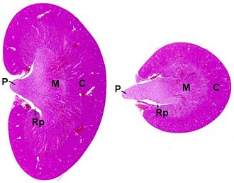
 Kidneys (P: papilla, M: medulla, C: cortex, Rp: renal pelvis). |
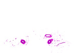
 Ureters. |
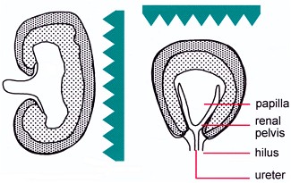
The transverse section of the kidney from the middle portion allows optimal examination of the renal pelvis, renal papilla and the junction with the ureter. The longitudinal section permits histological evaluation of a relatively large area of tissue which includes both renal poles. This is advantageous for the evaluation of any focal lesions. In addition, the regions of the renal pelvis close to the poles are of interest with respect to concretions and urothelial changes.
A slightly paramedian cut at trimming is helpful to get the full length of the renal papilla in section.
The ureter can be fixed on a cardboard together with a small amount of attached adipose tissue.
See also:
Introduction
Bachmann S, Kriz W (1998) Anatomy, cytology, ultrastructure nephron and collecting duct structure in the kidney, rat. In: Jones TC, Hard GC, Mohr U (eds) Monographs on pathology of laboratory animals. Urinary system, 2nd edition. Springer, Berlin Heidelberg New York Tokyo, pp 3–36 | |
Bannasch P, Schacht U, Storch E (1974) Morphogenese und Mikromorphologie epithelialer Nierentumoren bei Nitrosomorpholin-vergifteten Ratten. I. Induktion und Histologie der Tumoren. Z Krebsforsch 81: 311–331 | |
Cohen SM, Wanibuchi H, Fukushima S (2002) Lower urinary tract. In: Haschek WM, Rousseaux CG, Wallig MA (eds) Handbook of toxicologic pathology, 2nd edition, Vol 2. Academic Press, San Diego New York Boston, pp 337–362 | |
Corman BJ, Owen RA (1992) Normal development, growth, and aging of the kidney. In: Mohr U, Dungworth DL, Capen CC (eds) Pathobiology of the aging rat. Vol I. ILSI Press, Washington, pp 195–209 | |
Frith CH, Townsend JW, Ayres PH (1998) Histology, ultrastructure, urinary tract, mouse. In: Jones TC, Hard GC, Mohr U (eds) Monographs on pathology of laboratory animals. Urinary system, 2nd edition. Springer, Berlin Heidelberg New York Tokyo, pp 319–322 | |
Khan KNM, Alden CL (2002) Kidney. In: Haschek WM, Rousseaux CG, Wallig MA (eds) Handbook of toxicologic pathology, 2nd edition, Vol 2. Academic Press, San Diego New York Boston, pp 255–336 | |
Liebelt AG (1998) Unique features of anatomy, histology and ultrastructure, kidney, mouse. In: Jones TC, Hard GC, Mohr U (eds) Monographs on pathology of laboratory animals. Urinary system, 2nd edition. Springer, Berlin Heidelberg New York Tokyo, pp 37–57 | |
Short BG, Goldstein RS (1992) Nonneoplastic lesions in the kidney. In: Mohr U, Dungworth DL, Capen CC (eds) Pathobiology of the aging rat. Vol I. ILSI Press, Washington, pp 212–225 |
|
Trm V 5.00 |
Reference: Morawietz G, Ruehl-Fehlert C, Kittel B, et al. (2004) Revised guides for organ sampling and trimming in rats and mice – Part 3. A joint publication of the RITA and NACAD groups. Exp Toxic Pathol 55: 433–449 |