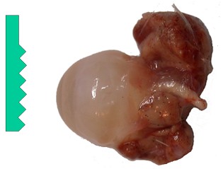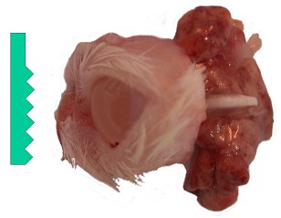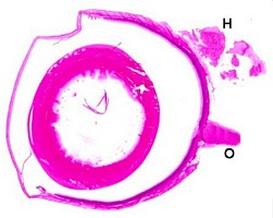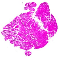
Eye, optic nerve and Harderian gland.
| Species: | Rats and Mice |
| Organs: | Eye Optic nerve Harderian gland Intraorbital lacrimal gland |
| Localization: | In the plane of the optic nerve |
| Number of sections: | 2 (1 per side) |
| Direction: | Longitudinal vertical |
| Remarks: | Optic nerve included. Optional: together with Harderian gland. Optional: together with eye lids for better orientation. For long-term studies, formalin fixation is generally sufficient. For other study types, fixation in Davidson's fixative is recommended to avoid detachment of the retina. |
 Eye, optic nerve and Harderian gland. |
 Eye. |
 Eye and eye lid. |
 Eye, Davidson's fixative, (O: optic nerve, H: Harderian gland) |
 Harderian gland. |
 Optic nerve, taken from the brain near to the optic chiasm. |
Each eye can be removed from the socket together with the optic nerve and the Harderian gland (optional) after fixation of the head. These organs are embedded in such a way that the section plane includes the anterior pole of the eye, the lens, the optic nerve and the gland.
Alternatively, each eye with the optic nerve can be taken at necropsy followed by the removal of the Harderian gland; in this case, both samples are fixed and embedded separately.
As an option, the optic nerve specimen can be taken at the base of the brain.
Relevant differences between rats and mice:
The rodent eye has no areas of increased visual acuity (fovea and macula), therefore orientation is not as critical as in the non-rodent eye. The Harderian gland lies intraorbitally and is cone-shaped in the rat and horseshoe-shaped in the mouse.
See also:
Salivary glands (extraorbital lacrimal glands)
Introduction
|
Trm V 5.00 |
Reference: Morawietz G, Ruehl-Fehlert C, Kittel B, et al. (2004) Revised guides for organ sampling and trimming in rats and mice – Part 3. A joint publication of the RITA and NACAD groups. Exp Toxic Pathol 55: 433–449 |