Revised guides for organ sampling and trimming in rats and mice: Parts 1 – 3
A joint publication of the RITA
*) and NACAD
**) groups
Note
The following text was mainly taken from part 1 of the
publication series and should provide a brief introduction. Where necessary, paragraphs from parts 2 or 3 were added. At the
end you will find some additional information regarding this Web site as well as a list of all
authors of the 3 parts.
Summary
This is the first part of a series of three articles on trimming instructions of rat and mouse protocol organs and tissues in regulatory type toxicity studies. It is based on the experience made in the European RITA and American NACAD working groups and is a continuation of trimming guides published in 1995 (BAHNEMANN et al.). The optimum localization for tissue preparation, the sample size, the direction of sectioning and the number of sections to be prepared is described organ by organ. These descriptions are illustrated for each organ by a schematic drawing and a macro-photograph showing the plane of section as well as a low power view of the H&E stained slide demonstrating the optimum "end-product".
This revision will improve the quality and efficiency of routine procedures and facilitate daily work in the histotechnical lab. It will also promote inter-study reproducibility and comparability and thus lead to a further improvement of the validity of historical control data.
Introduction
The first publication of the RITA group on the standardization of sampling and trimming procedures of organs in carcinogenicity studies was issued in 1995 (BAHNEMANN et al.). These guides were established based on the experience of pathologists and technicians from 20 pharmaceutical and/or chemical companies and research institutes in Europe working together in the RITA pathology data base project (MORAWIETZ et al. 1992, MORAWIETZ and RITTINGHAUSEN 1992, MOHR 1999, DESCHL et al. 2002). The primary goal of this approach was to standardize the laboratory techniques of tissue sampling and trimming procedures in terms of defining the sites at which samples should be taken, the amount of tissue which should be trimmed, the number of sections taken and the orientation of tissues on the slide. Beside the use of standardized nomenclature and diagnostic criteria (as also published and based on an initiative of the RITA group: MOHR 1992 - 1997, MOHR 2001), the application of standardized histology techniques is essential when comparing historical control data derived from different studies performed at different laboratories.
The 1995 paper (BAHNEMANN et al.) covered only the trimming of rat tissues, but was very positively received. With the kind permission of Urban & Fischer Verlag, that version of the RITA Trimming Guides has been available since 1998 free on the Internet (http://www.item.fraunhofer.de/reni/trimming). Also other publications (e.g. BONO et al. 2000) followed the basic criteria as outlined in the RITA paper.
In 1994 the North American Control Animal Database (NACAD) project was established and is operating in a way similar to RITA. In particular, the same data base structure is used, the data is stored on the same Fraunhofer ITEM data base server in Hannover, Germany, and NACAD is also based on standardized nomenclature and standardized diagnostic criteria (KEENAN et al. 2002). The companies involved in the NACAD project largely adapted their tissue trimming to the RITA trimming guides.
Although the initial idea was to standardize the trimming of tissues for carcinogenicity studies, the guides have also been successfully used for short term studies. Since different national or international guidelines require the processing of different protocol organs (LEBLANC 2000), we attempted to include the full set in this paper, knowing that not all organs are necessary for a particular study type.
Importance of standardization
The organs which must be routinely processed in a specific type of study (e.g. sub-chronic or carcinogenicity) are defined in various guidelines, regulating the approval/registration of pharmaceutical, chemical or agrochemical compounds. However, the guidelines usually do not mention which part of an organ should be examined histopathologically. This may result in poor comparability of incidence data obtained from different studies in different laboratories. Since the probability of detecting lesions is primarily related to the amount of the tissue examined, the need for standardization becomes clear. For larger organs (like lung or liver) it is necessary to define the number of sections and the specific lobe / area sectioned (e.g. left lateral lobe, right medial lobe of the liver). The cutting direction, either as a longitudinal or a transverse section, is in particular of importance for hollow organs (like the urinary bladder, uterus) in order to provide comparable areas of tissue for examination. Other technical procedures, such as instillation of fixative, decalcification, and the type of fixative used for particular organs influence the probability of detecting lesions in the final histological slide. A thorough understanding of the anatomic features (sub-sites) of all organs sampled (e.g. renal cortex and pelvis, adrenal cortex and medulla, seminiferous tubules and rete testis) is important to ensure an adequate histologic evaluation of all potential target sites in a given organ.
All these requirements were set in the frame of cost effectiveness, i.e. to gain a maximum of information with an acceptable investment of resources. It is not within the scope of this article to present sophisticated trimming procedures which may be required for specially designed mechanistic studies.
Revised and enhanced trimming guides
A number of reasons triggered the revision and enhancements of the criteria published in 1995. The main three are outlined below:
- In the last couple of years, a large number of mouse studies have been entered into the RITA and NACAD data bases, and the tissues have been histotechnically processed at the participating companies primarily following the guides established for the rat. The experience gained in the laboratories and the consideration of current literature showed that in some cases, an adaptation according to the anatomical situation in the mouse is necessary.
- The original (1995) trimming guides have been intensively discussed by the participants of the NACAD group and several modifications have been proposed, based on their practical experience. These proposals and suggestions for improvement were incorporated into the current paper, so that it now presents an international harmonization among both groups.
- The involvement of technicians in the information exchange stimulated the enhancements from a practical point of view. This resulted in the inclusion of macroscopic images of the organs and scans of the histological slides. Such histological images demonstrate how the final "product" should look, if the current guides have been applied.
Instructions and illustrations in individual organ guides
The revised and enhanced trimming guides are published in a series of three papers of which this is the first. The instructions are presented according to organ systems, organ-by-organ. In general, each organ description is valid for rat and mouse tissues but most of the gross and histopathological images are taken from the rat. If differences between the two species must be considered, they are mentioned in the text and/or in the figure legends.
For each organ the following information is usually presented:
- Localization: anatomical site or part of an organ from which a sample should be taken (i.e. lobe).
- Number of samples: number of organs (i.e. both for bilateral organs) or organ pieces prepared for evaluation (not necessarily identical with the number of slides / blocks).
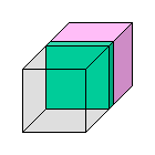
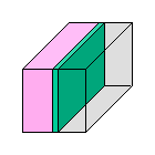
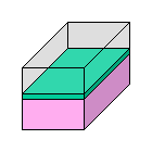
- Direction: direction (plane of section) in which an organ should be cut at trimming or microtome sectioning (see also the remarks at the end of this chapter). The proposed direction is shown in green color and optional sections (if defined) are shown in blue (see Fig. 1 for an explanation of the symbols used).
Fig. 1: Symbols used in the drawings and/or gross photographs to indicate the plane of section. a: cutting level parallel to the plane of the picture, b: cutting level perpendicular to the plane of the picture, c: cut level, 3-D
- Sample size: the size (area) of an organ or part of an organ which is sampled in a cassette for processing.
The sample size is determined by the size of an organ. For optimal fixation, sample thickness should not exceed 3–5 mm. In general, the examined area should be as large as possible and should contain the relevant anatomical structures. The tissue can be adapted to the size of cassettes by trimming the margins off.
- Optional remarks are used to present additional information, such as the instillation of fixative into the lung or the urinary bladder, optional recommended sections, placements of organs in cassettes, etc.
- Schematic drawings and/or gross photographs are given.
The plane of section is usually indicated in both images. Some of the gross photographs show the organ and trimming direction in situ. However, this is just for orientation purposes and it is recommended to remove the organ or tissue first. Trimming is performed as the next step, either on the fresh wet tissue or, in most cases, after fixation of the organ. Most of the gross photographs were taken from fresh unfixed organs. After fixation, tissue shrinkage and changes in color may lead to slight variations from the photos presented here.
- An image of a Hematoxylin and Eosin (H&E) stained slide is shown for the recommended section level (sometimes also for optional levels). Typical structures included in this section are indicated as necessary. As a routine, 10% buffered formalin (i.e., approx. 4% formaldehyde solution) is recommended as the fixative of choice. For some of the scans, the organs were fixed with Davidson's fluid. This is not indicated in the figure legend, since it usually does not influence the appearance of tissues at the low magnification used in the scans. If a special type of fixative is appropriate for a particular organ (e.g., eye or testis), it is mentioned in the organ manuscript.
- If helpful, images on histotechnical utilities (e.g. special cassettes, tools) are included.
- If appropriate, further information stating the reasons for specific sectioning levels or multiple sections, as recommended by the RITA/NACAD groups, is included.
- References to literature specific to a particular organ are included where appropriate and summarized at the end of each description.
In the descriptions the following terms are used for the determination of the trimming directions (see also Fig. 2 with a schematic presentation of the related cut levels):
- transverse: in a 90� angle to the long axis of an organ or part of an organ
- longitudinal vertical: in the direction of the long axis of the body, an organ or part of an organ in the dorsoventral axis
- longitudinal horizontal: in the direction of the long axis of the body, an organ or part of an organ, perpendicular to the dorsoventral axis
Fig. 2: Schematic presentation of the plane of section. a: transverse, b: longitudinal vertical, c: longitudinal horizontal
By defining either the "body", the "whole organ" or a "part of an organ" (for example a liver lobe or a certain part of the brain), as a unit of reference, it is relatively simple to precisely characterize a trimming direction by using only the three above defined terms and avoiding therefore the vast amount of anatomical terms and confusing synonyms present in literature. The schematic drawings and/or the gross images of the organs both include the trimming directions as colored lines or symbols to aid in orientation and identification of the correct sections.
Final technical remarks
Study types:
Although carcinogenicity studies in rodents are the focus of this publication, the same trimming procedures are also recommended for short-term rodent studies, unless specific target tissues or organs require an adaptation. In general, it is advisable to follow one standard trimming procedure in the laboratory for all studies to avoid technical inconsistencies and to facilitate inter-study comparison.
Additional organs:
Besides the organs addressed in these guides, special investigations or guidelines may require additional organs. Some can be found in the specimens described here, such as teeth in the transverse sections of the nasal cavity, brown adipose tissue at the renal hilus and heart base, white adipose tissue in the subcutis and the urethra of males in the prostate section. Others, however, may need adaptation of sampling procedures or collection of additional specimens.
Blocking:
The following recommendations should be taken into account:
- For reasons of economy it is desirable to have a small number of blocks per animal.
- Unduly large numbers of specimens in one block can lead to a loss of quality. Besides the obvious limitation of specimen size, difficulties may arise when cutting tissues with different physical properties in the same block.
- Small organs often benefit from being embedded by themselves which makes it easier to cut accurately the level of interest. Examples are the pituitary gland, the adrenal gland and the thyroid gland with parathyroid glands.
- Organs with similar cutting properties are often combined in one block.
- The pathologist benefits from being able to examine organs in functional groups, e.g. stomach together with intestine or a combination of the lymphoid organs in one block.
In order to reduce the number of slides, more than one block may be placed on a slide. Some laboratories see an advantage in collecting the adrenal glands, pituitary gland and thyroid gland with parathyroid glands on one slide which were processed in different blocks. An example of a blocking scheme reflecting the above mentioned considerations was given by K
RINKE (2000).
Number of sections:
The number of sections per specimen is usually one. If in some instances more than one section has to be taken, it should be borne in mind to evaluate the same amount of sections in all animals/groups to obtain comparable results.
Staining:
The H&E stain is regarded as the standard in toxicological studies. Other histological stains and immunohistochemistry can be applied as a routine or on a case by case basis in addition to the H&E stained sections.
Fixation:
In literature a volume ratio of tissue to fixative of 1:20 is often mentioned. However, much less fixative is sufficient, especially if a shaking device is used for freshly fixed tissues and/or fixative is replaced once. Tissues must be promptly and appropriately fixed by immersion. Adequate fixation time is necessary before tissue processing commences (C
RISSMAN et al. 2004).
Conclusion
The authors believe that this revision will improve the quality of routine procedures, facilitate daily work in the histotechnical lab and promote study comparability. It will also contribute to a further improvement of the validity of historical control data. As in the first paper (BAHNEMANN et al. 1995), main concerns taken into consideration were easy inter-study reproducibility and the relationship of cost and benefit. It is anticipated that these trimming guides will be made available on the Internet in a similar way as the first version.
Suggested reading
Besides the references mentioned in the individual organ guides, the authors suggest the following publications, if more or general information regarding anatomy, biology, histology or trimming of rodent tissues is needed. However, if chapters of these books are of particular interest for a certain organ guide, they are included in the related references.
A detailed anatomical description of the organ systems of the rat is given by HEBEL and STROMBERG (1986), BOORMAN et al. (1990) provide valuable information on the embryology, anatomy, histology and pathology of the Fischer rat, for some organs also with trimming proposals. For the mouse, comparable information can be found in the book by MARONPOT et al. (1999). Extensive information on normal anatomy, histology and physiology and their implications on toxicopathological aspects can be derived from the publications by KRINKE (2000) and HASCHEK et al. (2002).
Internet version
In addition to the data available in the printed publications, the Internet version provides the following features:
Navigation:
Organ manuscripts can easily be selected either from a list of organs arranged anatomically by organ systems (

button) or from an alphabetically sorted list (

button).
Images in larger size:
All images are available in a larger size: A click on an image within an organ manuscript displays this. Thumbnails on the right can be used for further selection. Clicking on the

button returns to the text display.
Interactive mode:
Organs, for which more than one cutting level is defined, can be viewed in the interactive mode (

button). If you hover with the mouse over the green or blue lines in the schemactical drawings and/or the macro images, the corresponding histological pictures are displayed. A click on a line shows the larger version of that image. Clicking on the
button returns to the text display.
Abstracts from PubMed:
As far as a literature reference is included in PubMed, a click on the

symbol shows the related abstract.
Regulatory guidelines:
At the bottom of each organ manuscript, a table shows, which guideline requires the processing of that organ. The sources from which this information was obtained are available on a
separate page.
Acknowledgements
The authors would like to thank Mr. THEODOR LAFORME and Mrs. BRIGITTE POSCHMANN (Bayer HealthCare AG) for the gross preparation of organs and histological slides; Mrs. PETRA HARTMANN, Mrs. MEIKE PETERSEN and Mrs. MARTINA NEUENHAUS (Bayer HealthCare AG) for scanning the histological slides, Dr. GERD-PETER FEHLERT for his help in image processing;
Mr. PETER KOCH and Mr. MIRCO JAHNKE (BASF AG) for the photo documentation on prostate preparation; Dr. MARTIN ROSENBRUCH (Bayer HealthCare AG), Dr. SUSANNE RITTINGHAUSEN and Dr. HEINRICH ERNST (Fraunhofer ITEM) for their scientific advice for organ preparation in inhalation studies;
Dr. WOLFGANG KAUFMANN (BASF AG) for his scientific advice regarding the nervous system, Dr. PAUL DESLEX (Pfizer Centre Recherche) for providing additional photographs and slides, Dr. THOMAS NOLTE (Boehringer Ingelheim Pharma GmbH & Co KG) and Dr. ANNE PROVENCHER BOLLIGER (Novartis Pharma AG) for help in establishing the bone marrow manuscript and Dr. MATTHEW JACOBSEN (Syngenta CTL) and Dr. RICHARD DOUGHTY (AstraZeneca) for their helpful input during the editing process.
References
 |
BAHNEMANN R, JACOBS M, KARBE E, et al.: RITA - Registry of Industrial Toxicology Animal-data - Guides for organ sampling and trimming procedures in rats. Exp Toxic Pathol 1995; 47: 247-266. |
|
BOORMAN GA, EUSTIS SL, ELWELL MR, et al. (eds): Pathology of the Fischer rat. Reference and atlas. Academic Press, San Diego, New York, London, 1990. |
|
BONO CD, ELWELL MR, ROGERS K: Necropsy techniques with standard collection and trimming of tissues. In: KRINKE GJ (ed) The laboratory rat. Academic Press, San Diego San Francisco New York, 2000; pp 569-600. |
|
CRISSMAN JW, GOODMAN DG, HILDEBRANDT PK, et al: Best practice guideline: toxicologic histopathology. Toxicol Pathol 2004; 32: 126-131. |
|
DESCHL U, KITTEL B, RITTINGHAUSEN S, et al.: The value of historical control data-scientific advantages for pathologists, industry and agencies. Toxicol Pathol 2002; 30: 80-87. |
|
HASCHEK WM, ROUSSEAUX CC, WALLIG MA (eds): Handbook of toxicologic pathology. 2nd edition, Academic Press San Francisco New York, 2002. |
|
HEBEL R, STROMBERG MW: Anatomy and embryology of the laboratory rat. BioMed, W�rthsee, 1986. |
|
KEENAN C, HUGHES-EARLE A, CASE M, et al.: The North American Control Animal Database: a resource based on standardized nomenclature and diagnostic criteria. Toxicol Pathol 2002; 30: 75-79. |
|
KRINKE GJ (ed): The laboratory rat. Academic Press, San Diego San Francisco New York, 2000. |
|
LEBLANC B: Pathology and tissue sampling protocols for rodent carcinogenicity studies; time for revision. Toxicol Pathol 2000; 28: 628-633. |
|
MARONPOT RR, BOORMAN GA, GAUL BW (eds): Pathology of the mouse. Reference and atlas. Cache River Press, Vienna, 1999. |
|
MOHR U (Editor): International classification of rodent tumours, Part 1: The rat. IARC Scientific Publications No. 122, Lyon, 1992 - 1997 (10 fascicles). |
|
MOHR U (Editorial): RITA – Registry of Industrial Toxicology Animal-data: Optimization of carcinogenicity bioassays. Exp Toxic Pathol 1999; 51: 461-475. |
|
MOHR U (Editor): International classification of rodent tumors, The Mouse. Springer, Berlin Heidelberg New York, 2001. |
|
MORAWIETZ G, RITTINGHAUSEN S: Variations in prevalence of endocrine tumors among different colonies of rats? A retrospective study in the Hannover Tumor REGISTRY Data Base. Arch Toxicol 1992; Supl 15: 205-214. |
|
MORAWIETZ G, RITTINGHAUSEN S, MOHR U: RITA – Registry of Industrial Toxicology Animal-data – Progress of the working group. Exp Toxic Pathol 1992; 44: 301-309. |
Authors of part 1 – 3
|
AXEL BUBE [3]
Drug Safety Evaluation Pathology, Aventis Pharma Deutschland GmbH, Hattersheim, Germany
|
|
ULRICH DESCHL [1]
Department of Nonclinical Drug Safety, Boehringer Ingelheim Pharma GmbH & Co KG, Biberach, Germany
|
|
PAUL DESLEX [1]
Department of Pathology, Pfizer Centre Recherche, Amboise, France
|
|
MICHAEL R. ELWELL [2]
Department of Pathology, Pfizer, Inc., Groton, CT, USA
|
|
SABINE HALM [3]
Department of Toxicology/Pathology/Animal Care, Abbott GmbH Co. KG, Ludwigshafen, Germany
|
|
JÜRGEN HELLMANN [3]
Institute of Toxicology, Merck KGaA, Darmstadt, Germany
|
|
ANKE HEUSER [3]
Institute of Pathology and Toxicology, ALTANA Pharma AG, Hamburg, Germany
|
|
KEVIN KEANE [3]
Schering-Plough Research Institute, Lafayette, NJ, USA
|
|
CHARLOTTE KEENAN [1]
Department of Preclinical Research and Development, Adolor Corporation, Malvern, USA
|
|
JAN KLAPWIJK [2]
Department of Pathology, Pharmacia Italia S.p.A., Gruppo Pfizer Inc., Nerviano, Italy
|
|
BARBARA LENZ [2]
Department of Toxicology, Hoffmann-LaRoche AG, Basel, Switzerland
|
|
BIRGIT KITTEL [1,2,3]
Department of Product Safety, Regulations, Toxicology and Ecology, BASF AG, Ludwigshafen, Germany
|
|
CHARLES R. MAHRT [1]
Department of Preclinical Toxicology, Pharmacia Corporation, Kalamazoo, MI., USA
|
|
GERD MORAWIETZ [1,2,3]
Department of Information Technology and Databases, Fraunhofer Institute of Toxicology and Experimental Medicine, Hannover, Germany
|
|
THOMAS NOLTE [1]
Department of Nonclinical Drug Safety, Boehringer Ingelheim Pharma GmbH & Co KG, Biberach, Germany
|
|
M. GERARD O'SULLIVAN [2]
Department of Pathology/Toxicology, 3M Pharmaceuticals, St. Paul, MN, USA
|
|
MERVYN ROBINSON [1]
Department of Regulatory Toxicology, Syngenta CTL, Alderley Park, Macclesfield, England
|
|
DANIEL R. ROTH [2]
Department of Pathology, Novartis Pharma AG, Basel, Switzerland
|
|
CHRISTINE RUEHL-FEHLERT [1,2,3]
Department of Toxicologic Pathology, Bayer HealthCare AG, Wuppertal, Germany
|
|
BARRY P. STUART [1]
Toxicology, Pathology and Veterinary Services Department, Bayer CropScience, Stillwell, KS, USA
|
|
PETER F. WADSWORTH [2]
Department of Pathology, AstraZeneca UK, Alderley Park, Macclesfield, England
|
*) RITA: Registry of Industrial Toxicology Animal-data. Members:
Abbott GmbH & Co KG, Ludwigshafen, Germany
ALTANA Pharma AG, Hamburg, Germany
AstraZeneca, Södertälje, Sweden and Macclesfield, England
Aventis Pharma Deutschland GmbH, Hattersheim, Germany
BASF AG, Ludwigshafen, Germany
Bayer HealthCare AG, Wuppertal, Germany
Boehringer Ingelheim Pharma KG, Biberach, Germany
Fraunhofer Institute of Toxicology and Experimental Medicine, Hannover, Germany
Hoffman-LaRoche AG, Basel, Switzerland
Merck KGaA, Darmstadt, Germany
Novartis Pharma AG, Basel, Switzerland
Pfizer, Amboise, France
Pharmacia, Nerviano, Italy
Syngenta CTL, Macclesfield, England
**) NACAD: North American Control Animal Database. Members:
3M Corporate Toxicology, St. Paul, MN, USA
Adolor Corporation, Malvern, PA, USA
Bayer CropScience, Stillwell, KS, USA
Pfizer, Inc., Groton, CT, USA
Pfizer, Inc., Ann Arbor, MI, USA
Pharmacia, Inc., Kalamazoo, MI, USA
R.W. Johnson Pharmaceutical Research Institute, Spring House, PA, USA
Schering-Plough Research Institute, Lafayette, NJ, USA